Wednesday, February 23, 2011

Medulloblastoma


Medulloblastoma is a series of tumors found in the cerebella.  It is more common in children than adults.  It is the most common malignant tumor affecting children.  The tumors usually occur in the cerebellum and posterior fossa of the brain.  Medulloblastoma has been known to metastasize to the bone. It can also spread throughout the Central Nervous System.
A common complication of this tumor is hydrocephalus.  Since it spreads through the CSF it causes intracranial pressure.  Some treatments of the hydrocephalus compression include a ventricular shunt or an endoscopic third ventriculostomy.  (http://emedicine.medscape.com)
 
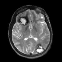
Axial CT image of lesion in posterior fossa

 
Sagittal MRI image showing medulloblastoma in cerebellum

Thursday, February 17, 2011

Cavernous Angioma

Cavernous Angioma
            Cavernous Angiomas (also called cavernous malformations) are developmental malformations of the vascular bed.  In other words, they are blood vessels that have formed abnormally or where they don't belong.  The malformation is known to leak blood and possibly cause small hemorrhages that do not usually amount to anything.  They are known to enlarge slowly over time as a result of the small hemorrhages.
            Most patients who have a cavernous angioma do not know they have one because they are asymptomatic.  Some possible symptoms include headaches and seizures. More serious symptoms occurring if the patient has a large hemorrhage are numbness, weakness, and vision changes depending on the location of the cavernous malformation.
            Treatment involves either observation or surgery if treatment is a possibility.  Most cases are minor and do not need treatment.  The more severe cases will undergo surgery if the location is easy to reach.  Other cases can undergo observation by getting annual MRI exams.
            MRI is the preferred exam to visualize a cavernous angioma.  CT is not the first choice because of the lack of specificity.  Particularly in MRI, a T2 weighted gradient echo sequence should be performed to find lesions that may not be seen on a standard spin echo sequence. 

Gradient echo axial MRI image showing multiple cavernous angiomas:

 Can you name the locations?  :)